
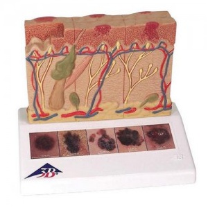
A. 특 징 (Features)
ㆍ병리학적인 피부 모형, 8배 확대
ㆍ건강한 피부와 대비되는 5가지 악성 흑색종 표현
- 건강한 피부
- 표피에 한정된 피부 표면의 암 세포
- 표피에 암 세포가 퍼져 있으며, 일부가 유두층에 침습
- 유두층에 암세포가 퍼짐
- 망상층에 암세포가 침습
- 위성 세포가 정맥과 접촉
B. 규 격 (Specifications)
14 x 10 x 11.5 cm; 0.2 kg
This 3B Scientific? Skin Pathology model shows healthy skin and 5 different stages of malignant melanoma on the front and back, enlarged 8 times:
- healthy
- malignant cells are found at the surface, within the epidermis
- malignant cells fill the epidermis, a few invade the papillary layer
- malignant cells fill the papillary layer
- malignant cells invade the reticular layer
- malignant cells have reached the subcutaneous fatty tissue, satellite cells approach a vein
In the top view of the skin cancer model, the individual stages of externally visible skin changes are shown, allowing for an assessment according to the “ABCDE” criteria. The sides of the skin cancer model show the various levels of invasion into the skin layers according to Clark (I-V) and the tumor thickness according to Breslow (in mm). 5 original color illustrations on the base of the skin cancer model show various types of malignant melanomas. The skin cancer model comes mounted on a base.
The skin cancer model is a great tool for illustrating this skin pathology.
* 계산서 발행 요청시 발행 가능합니다 . (카드 구입시 제외 )
* 무단 복재를 금합니다 .
상품정보고시
| 제품명 |
피부암 모형, J15 |
| 판매가격 |
가격문의 |
| 브랜드 |
3B Scientific |
| 원산지 |
EU |