¿À´Ã º» »óÇ° (0)
    no
    show
    °ßÀû¹®ÀÇ
    ½Ç½Ã°£°ßÀû
    ±â¼úÀÚ·á½Ç
    °øÁö»çÇ×
    Áú¹®°ú ´äº¯
    °í°´¼¾ÅÍ
    T.032-579-1191
    ¿ù - ±Ý : 09:00 ~ 18:00
    Á¡½É : 12:00 ~ 13:00
    Åä, ÀÏ, °øÈÞÀÏ ÈÞ¹«
    °í°´¼¾ÅÍ ¿¬°áÀÌ Àß µÇÁö¾ÊÀ»½Ã
    Q&A °Ô½ÃÆÇÀ» ÀÌ¿ëÇØÁÖ¼¼¿ä.

    ÇÇºÎ¾Ï ¸ðÇü, J15
    Skin Cancer Model
    ÆǸŰ¡°Ý °¡°Ý¹®ÀÇ
    ¿ø»êÁö EU
    ºê·£µå 3B Scientific
    Á¦Ç°ÄÚµå J15
    ÁÖ¹®¼ö·®
    »ó¼¼¼³¸í »óÇ°Èıâ(0) »óÇ°¹®ÀÇ(0) ±³È¯ ¹× ¹ÝÇ°/¹è¼Û/°áÁ¦


    4.JPG


    1.jpg


    A. Ư ¡ (Features)


    ¤ýº´¸®ÇÐÀûÀÎ ÇǺΠ¸ðÇü, 8¹è È®´ë


    ¤ý°Ç°­ÇÑ ÇÇºÎ¿Í ´ëºñµÇ´Â 5°¡Áö ¾Ç¼º Èæ»öÁ¾ Ç¥Çö


    - °Ç°­ÇÑ ÇǺÎ


    - Ç¥ÇÇ¿¡ ÇÑÁ¤µÈ ÇǺΠǥ¸éÀÇ ¾Ï ¼¼Æ÷


    - Ç¥ÇÇ¿¡ ¾Ï ¼¼Æ÷°¡ ÆÛÁ® ÀÖÀ¸¸ç, ÀϺΰ¡ À¯µÎÃþ¿¡ ħ½À


    - À¯µÎÃþ¿¡ ¾Ï¼¼Æ÷°¡ ÆÛÁü


    - ¸Á»óÃþ¿¡ ¾Ï¼¼Æ÷°¡ ħ½À


    - À§¼º ¼¼Æ÷°¡ Á¤¸Æ°ú Á¢ÃË


    B. ±Ô °Ý (Specifications)


    14 x 10 x 11.5 cm; 0.2 kg


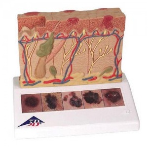
    This 3B Scientific? Skin Pathology model shows healthy skin and 5 different stages of malignant melanoma on the front and back, enlarged 8 times:


    - healthy


    - malignant cells are found at the surface, within the epidermis


    - malignant cells fill the epidermis, a few invade the papillary layer


    - malignant cells fill the papillary layer


    - malignant cells invade the reticular layer


    - malignant cells have reached the subcutaneous fatty tissue, satellite cells approach a vein


    In the top view of the skin cancer model, the individual stages of externally visible skin changes are shown, allowing for an assessment according to the ¡°ABCDE¡± criteria. The sides of the skin cancer model show the various levels of invasion into the skin layers according to Clark (I-V) and the tumor thickness according to Breslow (in mm). 5 original color illustrations on the base of the skin cancer model show various types of malignant melanomas. The skin cancer model comes mounted on a base.


    The skin cancer model is a great tool for illustrating this skin pathology.


    * °è»ê¼­ ¹ßÇà ¿äû½Ã ¹ßÇà °¡´ÉÇÕ´Ï´Ù . (Ä«µå ±¸ÀԽà Á¦¿Ü ) * ¹«´Ü º¹À縦 ±ÝÇÕ´Ï´Ù .
    »óÇ°Á¤º¸°í½Ã
    Á¦Ç°¸í ÇÇºÎ¾Ï ¸ðÇü, J15
    ÆǸŰ¡°Ý °¡°Ý¹®ÀÇ
    ºê·£µå 3B Scientific
    ¿ø»êÁö EU
    °ü·Ã»óÇ°
        
    »ó¼¼¼³¸í »óÇ°Èıâ(0) »óÇ°¹®ÀÇ(0) ±³È¯ ¹× ¹ÝÇ°/¹è¼Û/°áÁ¦
    ¹øÈ£ Á¦¸ñ À̸§ º°Á¡ ³¯Â¥
    ¾ÆÁ÷ ÀÛ¼ºµÈ »óÇ°ÆòÀÌ ¾ø½À´Ï´Ù.
     
    »ó¼¼¼³¸í »óÇ°Èıâ(0) »óÇ°¹®ÀÇ(0) ±³È¯ ¹× ¹ÝÇ°/¹è¼Û/°áÁ¦
    ¹øÈ£ Á¦¸ñ À̸§ ³¯Â¥
    ÀÛ¼ºµÈ »óÇ°¹®ÀÇ°¡ ¾ø½À´Ï´Ù.
      »óÇ°¹®ÀÇÇϱâ
    »ó¼¼¼³¸í »óÇ°Èıâ(0) »óÇ°¹®ÀÇ(0) ±³È¯ ¹× ¹ÝÇ°/¹è¼Û/°áÁ¦
    ¹ÝÇ°±³È¯
    ±¸ÀÔÁ¦Ç°ÀÇ ÀÌ»óÀÌ ÀÖÀ» °æ¿ì
    - ±¸ÀÔÈÄ 7ÀÏ À̳»¿¡ µ¿ÀÏÁ¦Ç°À¸·Î ±³È¯ °¡´ÉÇÏ¸ç ¿î¼Ûºñ´Â ÆǸÅÀÚ ºÎ´ãÀÔ´Ï´Ù.
    - ´Ù¸¥ Á¦Ç°À¸·Î ±³È¯, ¶Ç´Â ÀÌ»óÀÌ ¾ø´Â Á¦Ç°°ú ÇÔ²² ±³È¯À» ¿øÇÏ½Ç °æ¿ì ±¸¸ÅÀÚ²²¼­ ¿î¼Ûºñ¸¦ ºÎ´ãÇÕ´Ï´Ù.

    ±¸ÀÔÁ¦Ç°ÀÇ ÀÌ»óÀÌ ÀÖÀ» °æ¿ì (»ö»ó,»çÀÌÁî,´Ù¸¥»óÇ°±³È¯)
    - ±¸ÀÔÈÄ 7ÀÏÀ̳» ±³È¯ °¡´ÉÇÏ¸ç ±¸¸ÅÀÚ²²¼­ ¿î¼Ûºñ¸¦ ºÎ´ãÇÕ´Ï´Ù.

    !! ÁÖÀÇ»çÇ×
    ºñ´ÒÆ÷Àå ¹× TagÀÇ Æó±â ¶Ç´Â ÈÑ¼Õ µîÀ¸·Î »óÇ° °¡Ä¡°¡ ¸ê½ÇµÈ °æ¿ì¿¡´Â Á¦ÇÑ.
    ¹ÝÇ°½Ã¿¡ ÇØ´ç »çÀºÇ°ÀÌ ÀÖÀ» °æ¿ì °°ÀÌ º¸³»ÁÖ¼Å¾ß ÇÕ´Ï´Ù.
    ¹è¼Û¾È³»
    °áÁ¦ÈÄ 3~7ÀÏ À̳»¿¡ »óÇ°À» ¹Þ¾Æ º¸½Ç ¼ö ÀÖ½À´Ï´Ù.
    ±¹³» ÃÖ´ëÀÇ ¹°·ù»ç ¿»·Î¿ìĸÅù踦 ÅëÇÏ¿© ½Å¼ÓÇÏ°í ¾ÈÀüÇÏ°Ô ¹è¼ÛµË´Ï´Ù.
    ¸ðµç Á¦Ç°Àº ÂøºÒ ¹ß¼ÛµË´Ï´Ù. ´Ü 30¸¸¿ø ÀÌ»ó ±¸ÀԽà ¹«·á¹è¼ÛÀÔ´Ï´Ù.
    (Á¦ÁÖµµ¸¦ Æ÷ÇÔÇÑ µµ¼­,»ê°£Áö¿ªÀº Ç×°ø·á ¶Ç´Â µµ¼±·á°¡ Ãß°¡µË´Ï´Ù.)
    °áÁ¦¾È³»
    °áÁ¦¹æ¹ýÀº ½Å¿ëÄ«µå, ±¹¹Î/BC(ISP), ¹«ÅëÀåÀÔ±Ý, Àû¸³±ÝÀÌ ÀÖ½À´Ï´Ù.
    Á¤»óÀûÀÌÁö ¸øÇÑ °áÁ¦·Î ÀÎÇÑ ÁÖ¹®À¸·Î ÆÇ´ÜµÉ ¶§´Â ÀÓÀÇ·Î ¹è¼ÛÀÌ º¸·ùµÇ°Å³ª,ÁÖ¹®ÀÌ Ãë¼ÒµÉ ¼ö ÀÖ½À´Ï´Ù.
    »óÈ£ : ¿¡À̺ñÅ¥     ÁÖ¼Ò : ÀÎõ °è¾ç±¸ ÀÛÀüµ¿ 859-11     ´ëÇ¥ÀÚ : ÇãÁ¾¼ø     »ç¾÷ÀÚµî·Ï¹øÈ£ : 573-06-02527 (Á¤º¸È®ÀÎ)
    Åë½ÅÆǸž÷½Å°í : Á¦2022-ÀÎõ°è¾ç-1264È£     ºÎ°¡Åë½Å»ç¾÷ÀÚ¹øÈ£ :      °³ÀÎÁ¤º¸°ü¸®Ã¥ÀÓÀÚ : ±èÈñÁ¤     °³ÀÎÁ¤º¸º¸È£±â°£ : ȸ¿øÅ»Åð½Ã
    È£½ºÆü­ºñ½ºÁ¦°ø¾÷ü : µðÁöÀ¥